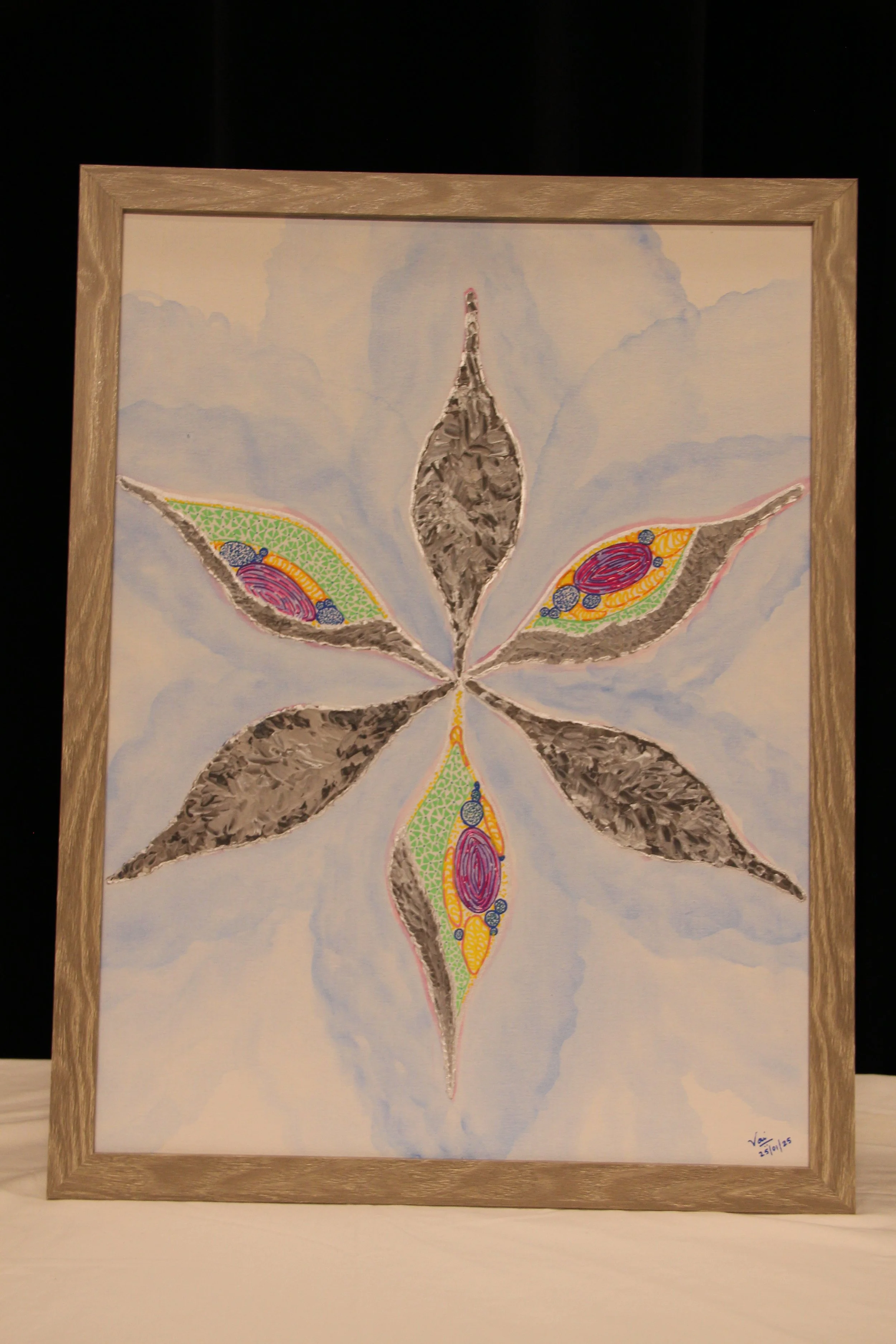
Under the electron microscope
Year : 2024
Media : Impasto, water and acrylic colours
Price : $150
An interpretive illustration, Under the Electron Microscope, showcases the imaging of the model diatom, Phaeodactylum tricornutum (PT). It is an 18 x 24 canvas illustration that depictAn interpretive illustration, Under the Electron Microscope, showcases the imaging of the model diatom, Phaeodactylum tricornutum (PT). It is an 18 x 24 canvas illustration that depicts the lead author's view of the diatom at 50,000 times magnification. Diatoms are a special class of algae with silica cell walls (commonly known as quartz, a glass-like substance), which make up 40% of the phytoplankton abundance in the ocean. The lead author has demonstrated different types of electron microscopy imaging techniques, including scanning electron microscopy (SEM) and transmission electron microscopy (TEM). Electron microscopy uses electron beams to create magnified images. an electron is a negatively charged particle that orbits a very small particle called an atom, similar to how planets orbit the sun. SEM helps image the exterior surfaces of the sample, whereas TEM creates images by passing electrons through the samples. In our interpretive illustration, the lead author utilized SEM in the scientific study and interpreted the TEM for the interior organelle structure (the colored internal organs of the diatom) of the PT based on the current understanding of scientific literature. Zentangle art was used to create the abstract shapes of the organelles present in the diatom.
